MRI
(Magnetic Resonance Imaging)
What is MRI?
MRI (Magnetic Resonance Imaging) is an imaging technique that can give very valuable information on a variety of structures in the body, which cannot be visualised with other imaging techniques such as X-ray’s and ultrasound. Think about very detailed images of the brain and spinal marrow, as well as cartilage, bone, various abdominal organs, ligaments and tendons in and around joints. Furthermore, by using MRI, the difference between normal and diseased tissue is very clear and all of this without the use of harmful radiation (such as by X-ray and CT scans)!
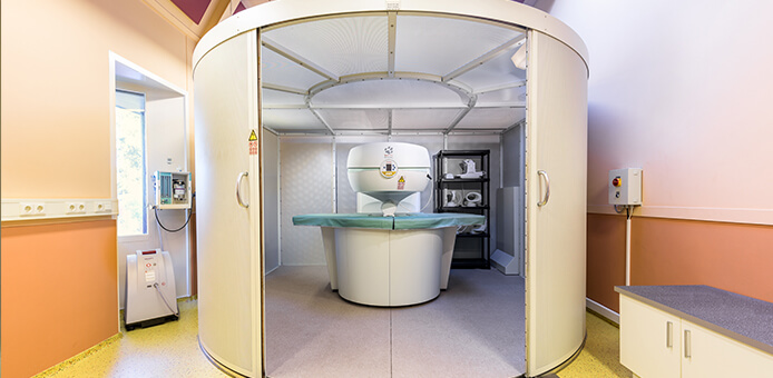
Simply put, an MRI scanner operates as follows:
The animal is placed in a strong magnetic field. As a result, all of the hydrogen nuclei (which, in fact are themselves just very small magnets) in the body stand in the same direction. Using a radio-frequency pulse, for a short moment the nuclei are flipped from that direction, whereupon they directly fall back into the same direction. Doing this, creates a radio sign. This is picked up by a coil-shaped antenna around the body and translated by computer into a picture. The rate at which the hydrogen nuclei fall back into their starting position is tissue-dependent, and creates contrast differences between tissues, which help us to distinguish the tissues from one another. It’s as if the body is three-dimensionally sliced into pieces by taking a large number of images of the part of the body that needs to be examined. All these images provide a wealth of information that is only visible using this method. The MRI is an entirely innocuous research technique for use on humans and animals alike.
Safety
Although the MRI technique is harmless to humans and animals, there are some safety issues that must be followed:
The MRI scanner contains a very strong magnet and therefore you must avoid contact with the following items: watches (stop immediately), bank- or credit cards (immediately erased), implants in your body (Pacemakers, insulin pumps, artificial joints etc.) and certain hearing aids.
In short, you are not allowed to enter the room where the MRI is situated if you are carrying any form of metal object on or with you. You will be asked to wait outside in the waiting room where you can enjoy a cup of coffee.
Why MRI?
Every owner who visits the vet with a sick animal is basically only interested in three things:
- What’s wrong with him? (Diagnosis)
- What are the chances of recovery? (Prognosis)
- What can be done? (Therapy)
It’s clear that you cannot answer questions two and three, if there is no diagnosis.
Often we cannot come to a clear diagnosis using conventional research techniques such as X-rays and ultrasound. Damage to ligaments, tendons, but also disorders of the brain and spinal cord are usually not visible on photos and ultrasound. In these cases, an MRI usually provides a diagnosis. Just like people with ‘unexplained’ complaints, a good diagnosis is sometimes only reached with the use of an MRI, it is only then that we can make a targeted course treatment and a good prognosis can be given!
Narcosis
If you have made an appointment for an MRI examination, your pet will need to be anesthetised. This is due to the fact that during the examination, which can take 45 minutes, he/she must not be allowed to move.
A good preparation for surgery under general anesthesia starts at home. The day before the MRI your pet may not eat any food after 18:00 hrs. Water is allowed! On the day itself please walk you dog before coming to the clinic at the appointed time. If you bring your pet to us, you do so under the (correct) assumption that he/she is surrounded by the best medical care. Therefore, in all cases, we perform a physical examination before the animal is put to sleep. There we pay special attention to the heart and lungs.
However, physical examination alone will not cover all possible problems. It may be necessary to perform a pre-anesthetic blood test. This may especially be necessary in older animals and animals with a reduced liver and kidney function. If another vet refers you to us, please bring the medical history of your pet with you (including any blood tests and or x-rays).
Just like medicine for humans, the anesthetics in veterinary medicine are very safe. For a healthy animal the risks of anesthesia are minimal. However, if there are any pre-existing health problems, anesthetic can cause certain problems.
If the pre-op search does not reveal any problems, our team can confidently bring your animal under anesthesia and begin with the examination. During the anesthesia the animal is monitored continuously.
After the investigation the animal is awakened, and is normally allowed to go home.
After the examination
Once the results of the animal study and all the images are received, we will then study the results before making a diagnosis. This diagnosis is often immediately made after the examination. If necessary, images can be additional submitted to experts in the field of medical imaging: Dr. I Gielen and Prof. H van Bree of the Rijksuniversiteit Gent.
If you are with us on a referral basis through your own vet, you will be referred back to them for the results and a treatment plan, we will of course send them a complete report of our findings.
You will receive a CD with all of the pictures from our research. We of course, retain all original data.
Syringomyelia
Chiari-like malformation-syringomyelia is a disorder that occurs in a small number of breeds. Best known is the condition in the Cavalier King Charles Spaniel, this is because a large number of Cavalier breeders have their spaniels tested for this disorder. However, it is undisputed that the disease occurs in a significant number of varieties, possibly even at a higher frequency than in the Cavalier King Charles Spaniel. For example breeds like chihuahua, petit griffon, King Charles spaniel.
The abnormality can only be diagnosed with the use of MRI. At present, an extensive screening program is running in order to map the spreading of CM/SM (Currently only the cavalier). By consistently screening for this deviation in recent years, a marked decline in the number of cases in Cavaliers with pedigree can be seen. Unfortunately, we do not see this in the so-called look-a-likes, these dogs are sold without pedigree as a Cavalier.
Dog-owners who want their dogs to be tested can arrange an appointment to screen for CM/SM. We do this at a greatly reduced rate. The images produced by us are judged by Dr. P.J.J. Mandigers, veterinary neurologist.